Examples of studies processed by the narnar app
Digital subtraction and color coding developed using our software contributed to the advancements in CEUS since its early years.
|
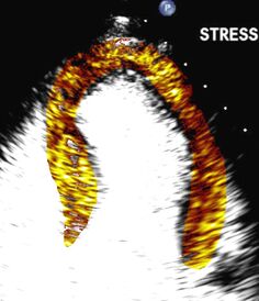
Perfusion Defect: High MI Imaging of AMI, alignment, averaging.
|
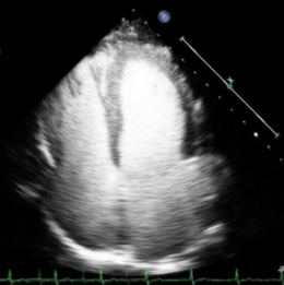
Averaging to improve myocardium delineation
|
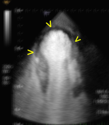
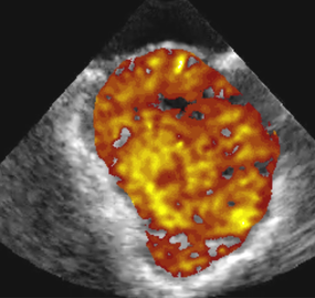
Normal Perfusion: Low MI Imaging and quantification of the heart
|
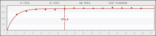
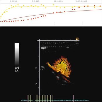
Quantification using time-videointensity replenishment curve and curve fitting to 1-exp function
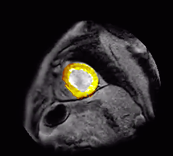
MRI: images can also be analyzed and quantified.
|
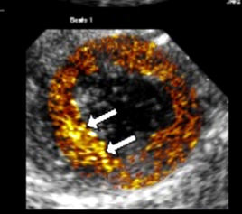
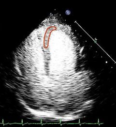
Activated Droplets: Infarct zone imaging
|
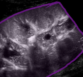
Kidney
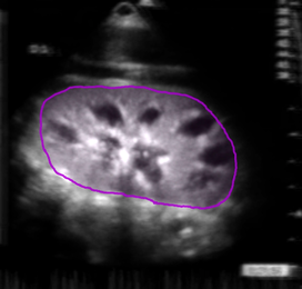
Normal kidney perfusion, images aligned and averaged.
|
|
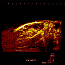
Liver
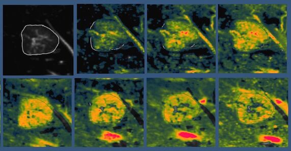
Focal nodular hyperplasia (FNH), a benign tumor-like mass of the liver.
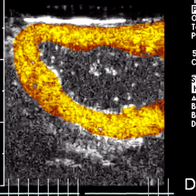
Placenta
Gradient of blood flow in a placental cotyledon.
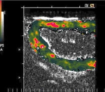
Uterus
Uterus (averaged)
|

a. Digitally subtracted, color coded image early during microbubble replenishment, uterus.
|
b. dtto, during the later microbubble replenishment phase
|
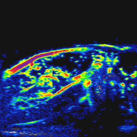
Uterus - parametric image of blood flow velocity.
|
Maximum Intensity Projection (MIP) images can also be rendered from a sequence of raw images.
|
MIP images are enhanced by color-coding.
|
Parametric image of blood flow in a small animal model.
© 2022 narnar, LLC. All Rights Reserved.